
Author: Coastal Ear, Nose & Throat

What Is Vestibular Migraine, and How Is It Different From Regular Migraines?

Vestibular Migraine

Migraines don’t always present as severe headaches; in fact, a vestibular migraine mainly affects balance, motion perception, and spatial orientation. Our ENT practice evaluates recurrent dizziness to diagnose whether a vestibular migraine or an alternative balance disorder is the primary cause. To find the right migraine treatment, it’s important to recognize the symptoms and communicate them to your doctor.

Vestibular vs. Regular Migraines: What’s the Difference?

While vestibular migraine and conventional migraine share an underlying neurovascular origin, their predominant clinical presentations diverge. Conventional migraine episodes standardly present with moderate to severe pulsating head pain that affects one side and worsens with activity. Associated features like nausea, pronounced light and sound sensitivity, and visual aura commonly accompany standard head pain.

In contrast, vestibular migraine mainly induces rotational vertigo, general dizziness, postural instability, and motion intolerance. Headache may occur, but it is not required during every vestibular episode. Flare-ups can feature an illusory sensation of internal or environmental spinning, head-movement-induced dizziness, and visual vertigo in crowded spaces.

Standard clinical diagnostic guidelines define vestibular episodes as lasting anywhere from five minutes up to 72 hours. Presentations frequently shift from episode to episode, which can obscure the underlying connection to migraine disease.

What Disorders Cause Vestibular Migraines?

Medical science recognizes that no single underlying disorder functions as the sole cause of vestibular migraine. The pathology seems to stem from shared central neural networks that process both vascular migraine signals and peripheral balance inputs. Genetic predisposition to migraines combined with a documented history of motion sensitivity markedly heightens susceptibility.

Typical episode triggers encompass the following:

  • Fluctuating sleep cycles or significant sleep deficits
  • Missed meals, fluctuating blood glucose, or systemic dehydration
  • Severe emotional stress or the physical transition period immediately following chronic stress
  • Menstrual cycle fluctuations or other physiological endocrine changes
  • High-contrast visual patterns, strobe effects, or glaring light environments
  • Prolonged travel, turbulent physical motion, or rapid digital screen scrolling
  • Alcohol intake, fermented foods, cured meats, or idiosyncratic dietary sensitivities
  • Ambient weather shifts, storm fronts, or crowded environments causing sensory overload

What Are the Symptoms of Vestibular vs. Regular Migraines?

Typical migraine attacks are often recognized by headache, but migraine symptoms extend beyond pain. A person may experience nausea, vomiting, visual aura, fatigue, brain fog, neck discomfort, or sensitivity to light, sound, and smell.

Symptoms associated with vestibular migraine often encompass:

  • True rotational vertigo or generalized non-rotational vertigo sensations
  • Persistent postural unsteadiness, gait instability, or loss of balance
  • Cervicogenic or vestibular dizziness exacerbated by head motion
  • Gastrointestinal nausea provoked by physical movement or riding in vehicles
  • Sensory intolerance triggered by scrolling monitors, bustling public crowds, oncoming traffic, or geometric lines
  • A sensation of rocking, tilting, floating, or being pulled
  • Heightened photophobia, phonophobia, or generalized visual sensitivity
  • Diffuse head pressure, orbital tension, or characteristic throbbing migraine pain
  • Temporary ear pressure, fullness, or ringing

The clinical timeframe, duration of dizziness, and identified triggers are vital for distinguishing vestibular migraine from other vestibular etiologies. Newly developing dizziness linked to facial numbness, extremity weakness, speech impairment, double vision, syncope, or an inability to walk demands immediate emergency attention.

The Coexistence of Vestibular Migraine and Typical Migraine Episodes

Yes. True rotational vertigo and classic migraine head pain can develop within the identical clinical attack. Dizziness might manifest as a premonitory symptom before head pain, peak while the headache is active, endure following pain resolution, or strike alone. Over years of disease progression, individuals frequently oscillate between headache-heavy attacks and balance-centric vestibular events.

An episode of vertigo does not have to feature head pain to be classified within the migraine spectrum. Maintaining a dedicated symptom journal helps track chronological correlations that are easily overlooked during clinical consultations. Record the timing and duration of dizziness, headache features, sensory sensitivity, menstrual cycle, sleep, meals, possible triggers, medications, and response to treatment. These objective records provide invaluable diagnostic clarity during your comprehensive evaluation.

Diagnostic Workup and Clinical Criteria for Vestibular Migraines

Diagnosing vestibular migraine cannot be accomplished via a single diagnostic blood test, radiologic scan, or balance assessment. Diagnostic evaluation initiates with a structured examination of symptom chronology and a confirmed history of migraine with or without aura. Consensus clinical guidelines specify at least five moderate to severe vestibular episodes spanning five minutes to 72 hours, accompanied by migraine characteristics in at least half of the events.

We execute an exhaustive diagnostic series involving neurological testing, videonystagmography, direct ear examination, hearing testing, and balance assessments. Tailored to your clinical needs, diagnostic protocols can incorporate pure-tone audiograms, positional testing, specialized vestibular studies, or MRI scans. Such objective tests are crucial to differentiate vestibular migraine from other balance disorders presenting with similar symptoms. A formal diagnosis of vestibular migraine is established only when clinical criteria are satisfied and no alternative condition provides a better explanation.

Treatment Strategies: Managing Vestibular Migraines Effectively

Treatment is individualized according to attack frequency, severity, associated headache, other health conditions, and medication risks. Most individuals find the greatest relief through an integrated regimen combining daily preventive protocols with an acute rescue plan.

We help build a customized treatment plan, which may include:

  • Establishing structured daily routines for sleep, timely meals, fluid intake, and low-impact cardiovascular exercise
  • Isolating and minimizing contact with consistent environmental or dietary triggers
  • Implementing evidence-based stress-reduction protocols, biofeedback, or behavioral therapies
  • Prescribing fast-acting antiemetics to manage acute nausea and autonomic symptoms
  • Measured, temporary use of vestibular suppressant pharmaceuticals during severe vertigo crises
  • Prophylactic regimens involving beta-blockers, calcium channel blockers, topiramate, tricyclic antidepressants, or CGRP inhibitors
  • Customized vestibular rehabilitation therapy to address persistent disequilibrium and visual motion intolerance

Prescription vestibular suppressants must be used with caution, as continuous dosing induces significant sedation and inhibits central vestibular compensation. Medication choices should be made with us rather than borrowed from another person’s migraine plan. Achieving optimal symptom control typically demands careful dose titration and continuous symptom tracking over time.

Request a Clinical Evaluation for Vestibular Migraine

If ongoing vertigo attacks, motion hypersensitivity, or gait instability compromise your daily functioning, schedule an assessment with our clinic today. Your comprehensive consultation entails a thorough evaluation of migraine history, medical background, and individual goals, using targeted diagnostics to isolate vestibular migraine from BPPV, Ménière’s disease, or other vestibular etiologies. Get in touch with our ENT clinic now to request your vestibular migraine appointment.

Understanding Tonsil Stones: Causes, Symptoms, and Effective Removal

Tonsil Stones

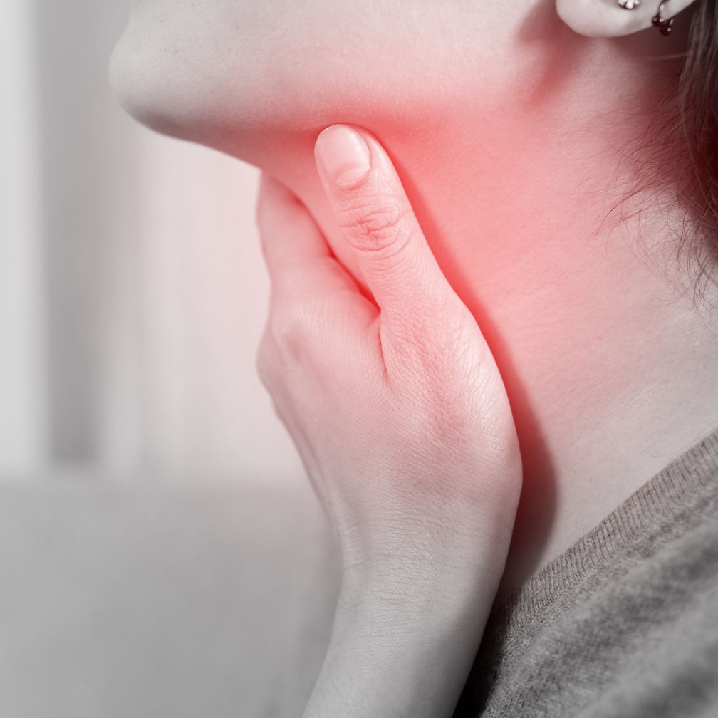
Tonsil stones consist of condensed, hardened accumulations that often drive annoying and ongoing throat symptoms when neglected. Our specialized medical team guides patients through safe home protocols and provides clinical treatments when tonsil stone symptoms persist. Familiarizing yourself with tonsil stone etiology and symptom management determines whether you require professional removal or advanced treatment.

What Causes Tonsil Stones?

Tonsil stones, or tonsilloliths, form inside the irregular surface depressions and crevices termed tonsillar crypts. Cellular debris, microscopic food particulate, salivary proteins, oral mucus, and proliferating bacteria aggregate inside these tissue pockets. With continuous mineralization, the condensed debris hardens into a firm, whitish-yellow pebble-like formation.

Some people naturally have deeper or more numerous crypts. Chronic infectious episodes leave behind scarred, expanded tonsil pockets that readily trap food particles and bacteria. Supplementary factors driving tonsil stone formation include:

  • Ongoing, repetitive bouts of infectious tonsillitis
  • Hypertrophic, heavily creased tonsillar architecture
  • Systemic dehydration and inadequate salivary flow (xerostomia)
  • Postnasal drainage and excess mucus
  • Irregular oral hygiene routines, poor flossing, or skipped tongue brushing
  • Cigarette use, toxic inhalation, or ongoing oral mucosal irritation

Tonsilloliths cannot be transmitted to other individuals and do not automatically reflect inadequate oral hygiene.

How to Identify Tonsil Stone Symptoms

Small stones may cause no symptoms and be noticed only during a dental or throat examination. When symptoms occur, one of the most common complaints is persistent bad breath. Colonizing bacteria feeding on stagnant debris generate distinct volatile sulfur compounds that cause bad breath and foul taste.

Additional documented clinical symptoms include:

  • White or yellow pebble-like material on a tonsil
  • Chronic pharyngeal irritation, throat scratchiness, or localized soreness
  • The sensation of something lodged in the throat
  • Dysphagia (difficulty swallowing) or odynophagia (painful swallowing)
  • Coughing or frequent throat clearing
  • Referred ear pain accompanied by entirely normal middle-ear findings
  • A swollen or tender tonsil
  • A lingering, noxious metallic or acrid taste along the tongue

It is crucial to distinguish benign tonsil stones from acute infectious tonsillitis. Tonsillitis presents as a microbial inflammation causing intensely red, enlarged tonsils, acute throat pain, fever, and adenopathy. White pharyngeal patches can stem from numerous underlying bacterial, fungal, or viral causes. An expert ENT evaluation in our clinic isolates tonsilloliths from acute viral, bacterial, or neoplastic throat diseases.

How Can I Treat Tonsil Stones at Home?

Asymptomatic, small tonsil stones can safely be left untreated. If a tonsil stone produces symptoms, implement gentle interventions that do not lacerate or damage the tonsil.

Clinically supported home protocols include:

  • Perform warm saline gargles consistently, particularly following meals.
  • Brush twice every day and floss regularly to reduce retained food particles and microbial loads.
  • Maintain optimal systemic hydration to prevent oral dryness and stimulate salivary flow rates.
  • Attempt soft gargling or gentle coughing to see if mechanical fluid shifts loosen the deposit.
  • Utilize an antimicrobial, non-alcoholic mouthwash if suggested by our clinical team.

We strongly advise consulting our clinical team before trying to manually dislodge any tonsil stone. You must never probe, scrape, or extract stones using fingernails, wooden toothpicks, metal instruments, or sharp items. Halt attempts at once should you experience localized bleeding, acute pain, or violent gagging.

Understanding Why Tonsilloliths Recur

Dislodging an active tonsil stone clears the blockage, yet the underlying pocket remains unchanged. Deep anatomical fissures will inevitably continue pooling oral secretions, food particles, microbes, and shed cells. That is precisely why individuals with irregular tonsillar surfaces or recurrent throat infections form stones despite great oral hygiene.

Rigorous home oral routines can limit bacterial colonization, but they cannot remodel anatomical tonsil crypts. Commit to consistent brushing, interdental flossing, tongue cleaning, optimal water intake, and post-prandial gargles. Address ongoing nasal symptoms or dry mouth with appropriate professional care. Repeated tonsil stones are seldom hazardous, yet intractable symptoms merit an ENT evaluation to weigh long-term solutions.

When to Consult an ENT Specialist for Tonsil Stones

Book an appointment with our office when at-home remedies fail to help or stones routinely degrade comfort, deglutition, or sleep. Our clinicians will confirm whether tonsilloliths are causing your symptoms and examine the throat for secondary infections.

You should seek focused clinical care for:

  • Persistent bad breath despite thorough oral and dental care
  • Dense, deeply seated tonsil stones or rapidly recurring debris formations
  • Chronic pharyngeal pain, unilateral ear discomfort, or progressive dysphagia
  • Localized hemorrhage or tissue laceration occurring after self-extraction efforts
  • Fever, pus, tender neck glands, or repeated throat infections
  • Marked tissue edema or significant asymmetry where one tonsil appears larger
  • Symptoms that continue even when no stone is visible

Critical pharyngeal symptoms must never be downplayed as uncomplicated tonsil stones. Seek urgent care for difficulty breathing, drooling, or inability to swallow liquids, rapidly increasing swelling, severe one-sided pain, or a muffled voice. These alarming signs often suggest complex infectious emergencies requiring immediate hospital-based intervention.

Professional Tonsillolith Extraction by an Otolaryngologist

Our team evaluates tonsil contours, logs recurrence history, and investigates whether symptoms stem from acute tonsillitis, postnasal drip, reflux disease, or oral pathology.

If a stone cannot be removed safely at home, we may dislodge it with appropriate instruments during an office visit. In addition, we design preventive oral health protocols, address contributing reflux or sinus disease, and prescribe antibiotics for active bacterial infections.

Undergoing a tonsillectomy surgically excises the palatine tonsils, permanently eliminating the tissue crypts where stones take shape. Given that operative tonsil removal carries risks associated with general anesthesia, post-procedure recovery, and bleeding, it is reserved for chronic or debilitating cases where benefits exceed surgical risks.

Arrange a Clinical Throat and Tonsil Evaluation

If persistent tonsil stones cause ongoing throat irritation or interfere with comfortable breathing and swallowing, contact us to schedule an exam. Through a thorough clinical exam, we can confirm if visible white material is indeed a tonsil stone while checking for localized inflammation or alternative pathologies.

Contact our ENT clinic today to request your appointment.

Why Q-Tips Harm Your Ears and How to Safely Remove Cerumen

Q-Tip

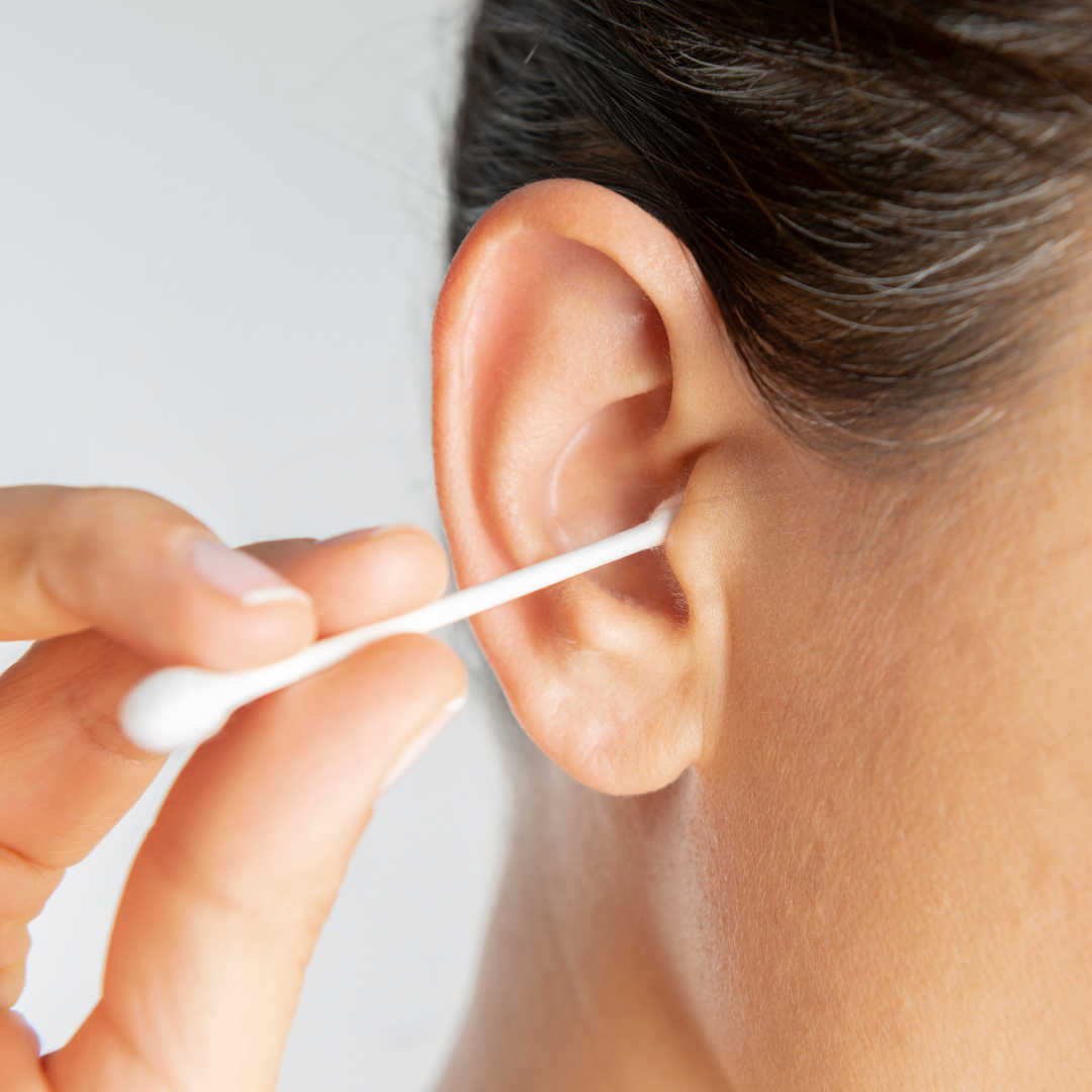
Even though cotton swabs seem practical for routine hygiene, probing the inner canal creates deeper impactions and threatens long-term ear health. We assess whether your earwax demands professional intervention and provide customized care strategies that match your clinical profile. Ears don’t always need manual cleaning; however, if wax builds up, we can help patients safely remove excess wax.

Clinical Reasons Why Q-Tips Damage the Ear Canal

Earwax, or cerumen, is not dirt. Cerumen collects particulate matter, inhibits microbial proliferation, moisturizes the epithelial lining, and shields sensitive canal tissues. Routine chewing and epithelial cell migration continuously transport spent wax toward the concha, where it falls out or clears during bathing.

A Q-Tip placed inside the canal can disrupt this self-cleaning process. Even if the swab gathers a trace of surface cerumen, it routinely compacts the remaining mass against the eardrum. Chronic swab insertion frequently compresses cerumen into a dense impaction, triggering a sensation of aural fullness that invites improper cleaning cycles.

Although swabs can sanitize the outer folds of the auricle, they should never penetrate the delicate ear canal.

Clinical Complications Associated with Q-Tip Insertion

The skin lining the ear canal is thin and easily scratched. Minor scratches can trigger localized pain, bleeding, and bacterial infiltration, substantially elevating the risk of otitis externa. The cotton end can also dislodge from the applicator, creating a retained foreign body within the ear.

Documented injuries associated with cotton swabs include:

  • Earwax compressed tightly against canal walls
  • Skin abrasions, tissue swelling, severe irritation, and active bleeding
  • Pathogenic infection involving the ear canal
  • Retained cotton tips or foreign debris inside the canal
  • Ruptured, torn, or perforated tympanic membrane
  • Dislocation or injury of the delicate hearing ossicles
  • Temporary conductive loss or permanent auditory changes
  • Persistent tinnitus, balance instability, or severe otalgia

Severe injuries are infrequent yet emerge readily if an arm is bumped, the swab slips, or the stick is driven too far.

How Often Should Earwax Get Removed?

Clinical extraction of earwax is not routinely required on a fixed schedule for most patients. Normal anatomical mechanisms steadily push cerumen toward the exterior, where regular washing rinses the visible concha. Overcleaning can dry or irritate the canal and may increase the likelihood of infection or impaction.

Intervention becomes necessary whenever wax creates an anatomical blockage, obstructs otoscopic examination, disrupts hearing aid operation, or causes aural distress.

What Are Safe Ways to Remove Earwax At Home?

When cerumen creates no clinical symptoms, leaving the canal alone remains the most prudent approach. Restrict hygiene to wiping the outer ear folds with a damp cloth while permitting the canal to self-evacuate.

When dry wax demands pre-treatment, we often advise using an OTC cerumenolytic solution or drops composed of glycerin, mineral oil, carbamide peroxide, or saline. Dispense only the directed amount and restrict use strictly to the timeframe specified on the product packaging. Discontinue the product right away if you encounter otalgia, severe dizziness, contact dermatitis, or aggravating symptoms.

Home irrigation is not appropriate for everyone and should only be attempted after a professional confirms it is safe and explains the technique. You must never employ high-pressure dental water jets for ear clearing. Avoid ear candles, metal scoops, camera-guided tools, hairpins, and home suction devices. These questionable products can burn sensitive tissues, lacerate the canal, rupture the eardrum, or compact wax deeper.

When Is Professional Earwax Extraction Necessary?

Schedule a professional visit if the canal remains occluded after softening treatments, cerumen appears deeply lodged, or home remedies fail. Seeking professional care is essential if a patient cannot stay still, exhibits narrow canal anatomy, wears hearing aids, or possesses medical risks that rule out drops or flushing.

Schedule an ear exam promptly if you experience:

  • Persistent otalgia, stubborn itching, or painful sensitivity around the ear
  • Hemorrhagic discharge, purulent fluid, clear otorrhea, or foul-smelling drainage
  • Fever or swelling around the ear
  • Sudden-onset tinnitus or vestibular dizziness
  • Sudden hearing drops or auditory deficits that remain after wax removal
  • Concern regarding a perforated eardrum or foreign cotton debris
  • Recurrent impaction that affects hearing devices

You should always treat sudden unilateral hearing loss as an urgent medical emergency instead of assuming simple cerumen occlusion. Early hearing testing easily separates a physical wax impaction from a critical inner ear condition.

In-Office Procedures for Clearing Earwax Blockages

We initiate the appointment by visualizing the canal through a diagnostic otoscope or microscope to verify wax presence and check for signs of infection, laceration, or eardrum tears. The safest removal method depends on wax consistency, canal anatomy, medical history, previous surgery, and the patient’s ability to tolerate the procedure.

Expert removal procedures include:

  • Topical cerumenolytic drops applied prior to or during the clinical visit
  • Regulated mechanical irrigation with body-temperature saline for sealed eardrums
  • Safe, gentle suction extraction executed under direct microscopic guidance
  • Manual removal with a loop, curette, or small forceps

Visual guidance empowers the clinician to protect delicate canal skin and pause immediately if sensitivity is detected. You may need more than one method for dry or tightly packed wax. Once the wax is cleared, we reexamine the ear to ensure the auditory canal and eardrum remain completely healthy. If aural fullness, ear ringing, dizziness, or auditory impairment remains after cleaning, further diagnostic evaluations can identify the root issue.

Request an Appointment for Clinical Earwax Removal

When your ears feel plugged or you notice auditory changes, schedule an appointment with our specialists instead of grabbing a cotton swab. Your scheduled visit includes reviewing active symptoms, past ear procedures, hearing aid status, home management methods, and individual risk factors for cerumen impaction. Contact our office directly to book your safe cerumen-removal appointment.

When to Take Your Child to an ENT Specialist: Essential Signs for Parents

Child seeing an ENT doctor

At what point should your child consult an ENT specialist? This concern is commonly voiced by families dealing with lingering earaches, stubborn sinus problems, or ongoing throat conditions. While general pediatric practitioners address basic childhood conditions, distinct complications necessitate intervention by an Ear, Nose, and Throat (ENT) specialist.

Warning Signs That Call for an ENT Specialist Evaluation

Pediatric patients typically recover quickly but remain susceptible to ordinary colds, seasonal flu, and sporadic pharyngitis. These basic medical issues are normally treated by local pediatricians. Nonetheless, when health complaints become continuous or recurrent, scheduling an assessment with an ENT specialist is necessary.

Below are specific situations where taking your child to an ENT specialist offers clear value.

Chronic ear infections

If your child experiences more than three ear infections within six months, it may indicate a chronic issue that requires specialized treatment. Experienced ENT specialists deliver targeted therapies to stop repeated infections while protecting vital auditory development.

Addressing Recurrent Strep Throat and Tonsillitis

Six or more episodes of tonsillitis or strep throat in a year suggest a recurring problem that might benefit from ENT evaluation. Otolaryngology doctors review symptom severity to judge if a tonsillectomy (removal of the infected tonsils) is appropriate to eliminate subsequent throat episodes.

Identifying Chronic Sinus Infections in Children

Experiencing three or more confirmed sinus infections within a year, highlighted by lingering facial soreness, cloudy nasal secretions, and continuous coughing, warrants advanced ENT assessment. Skilled physicians establish comprehensive protocols designed to resolve acute symptoms and inhibit subsequent sinus complications.

When Persistent Nosebleeds Require Clinical Care

Stubborn nasal bleeds showing no improvement with routine home care may signal localized vessel vulnerabilities that an ENT specialist addresses properly.

Navigating Childhood Allergies and Asthma Concerns

Children facing severe allergic rhinitis commonly endure sinus blockage, prolonged coughing, and labored inhalation, which worsen significantly when asthma is present. Skilled ENT clinicians align with pediatric allergists to establish complete and effective airway management protocols.

Understanding Signs of Pediatric Sleep Apnea

Heavy nighttime snoring among children occasionally signals underlying sleep apnea, a critical airway issue that demands review by a pediatric ENT specialist. These physicians comprehensively inspect pediatric airways to determine optimal therapies that support undisturbed nightly rest.

Additional Pediatric Otolaryngology Concerns

Diverse medical scenarios including gait imbalance, sudden hearing impairment, chronic dysphagia, and maxillofacial trauma remain central specializations for practicing ENT specialists. They have the expertise to diagnose and treat these issues, ensuring your child’s overall health and well-being.

Determining When to Seek Clinical Help

Whenever you detect sustained complaints including chronic coughing, respiratory compromise, recurrent illness cycles, or alarming ear, nose, and throat manifestations, arranging care with an ENT specialist is prudent. Swift diagnostic action averts progressive anatomical complications and noticeably improves your youngster’s developmental health.

Choosing the Best ENT Specialist for Your Child

Locating the best ENT specialist involves reviewing their credentials in pediatric otolaryngology, their individualized approach to care, and their personal communication standards. A distinguished physician treats the immediate physiological problem while delivering empathetic guidance and structural support across every recovery stage.

In review, while standard infectious bugs occur during early maturation, continuous or repetitive head and neck complaints justify a consultation with an ENT specialist. Their advanced clinical diagnostic skills and targeted procedural treatments deliver substantial advantages when protecting your youngster’s health. If you notice troubling issues involving your child’s ears, nose, or throat, contact an experienced ENT specialist promptly for comprehensive care.

What Causes an Unresolved Cough? Recognizing When to See a Physician

July ENT 3 Cough Go Away

A cough is usually just a temporary symptom, often linked to a cold, flu, or allergies. Under normal circumstances, the issue resolves naturally within several days. But what happens when the cough doesn’t go away? If you endure coughing day in and day out, it can represent something beyond a trivial annoyance. Sustained coughing might signify that a separate internal concern needs addressing. Consequently, how should you proceed if your coughing continues?

Questioning why your throat irritation refuses to clear up is entirely understandable. Whether it’s simply irritating or potentially serious, figuring out the cause is the first step toward finding relief.

Examining the broad impact of a sustained cough on your lifestyle

Any lingering cough that resists improvement may disrupt your life far beyond a minor annoyance. Even a modest, continuous cough carries the potential to disturb your regular schedule. Below are several primary ways a persistent cough impacts your well-being:

Social Stress: Within contemporary public settings, frequent coughing frequently invites immediate, uncomfortable looks. Observers often infer that you are sick, even when your coughing originates from non-infectious causes. This dynamic often results in uncomfortable settings as people distance themselves or shy away from engagement. Poor Sleep Quality: Sustained coughing easily ruins an individual’s overnight rest. Whether it stops you from drifting off or startles you awake throughout the night, a continuous cough limits functional recovery, producing daytime sluggishness and irritability. Impaired Nourishment: A lingering cough frequently interferes with the process of eating. The physical movement of coughing can impede smooth swallowing, occasionally sparking choking episodes or intensified coughing mid-meal. Over time, this dynamic reduces the pleasure of eating and can even precipitate weight loss.

Distinguishing the traits of a sustained cough

So, what factors divide a lingering cough from a basic viral illness? Overall duration functions as a primary differentiating factor. Standardly, coughing that continues for six to eight weeks or beyond is defined clinically as chronic. Yet duration does not act as the exclusive diagnostic variable. Additional clinical features often present alongside a chronic cough, including:

  • Upper airway secretions
  • Gastric acid reflux or heartburn
  • Continuous throat discomfort
  • Involuntary weight decline
  • Nocturnal diaphoresis
  • Coughing up bloody mucus
  • Dyspnea or respiratory wheezing
  • Vocal strain or hoarseness
  • Occasional fever

While these indicators range from mild to severe, encountering any combination warrants a medical evaluation to identify the root cause and initiate proper management.

Identifying the primary triggers of an ongoing cough

A chronic cough generally serves as a warning that an alternate medical process requires attention. Pinpointing the underlying trigger enables you and your clinician to structure a successful therapeutic approach. Here are some of the most common reasons for a chronic cough:

Pulmonary Disorders: Long-term lung conditions such as asthma, chronic obstructive pulmonary disease (COPD), or pulmonary fibrosis frequently induce chronic coughing. While asthma generally responds well to targeted inhalers and environmental adjustments, disorders such as COPD present greater complexity and can progress, driving increased coughing frequency. Allergic Triggers: Even though sneezing remains the most recognized allergic indicator, persistent coughing can also indicate an allergic reaction. If your cough is seasonal or linked to exposure to certain allergens, such as pollen or pet dander, allergies could be the underlying cause. Sinus Discomfort: Issues involving the paranasal sinuses, including bacterial infections or chronic sinusitis, often promote post-nasal discharge. The resulting mucus discharge irritates throat tissues and sparks a continuous coughing pattern. Based on clinical findings, management strategies range from conservative pharmaceutical options to surgical intervention for advanced cases. Digestive Disorders: Acid reflux and GERD (gastroesophageal reflux disease) can cause stomach acid to flow back into the esophagus, irritating your throat and leading to a cough. Resolving the gastrointestinal reflux issue frequently reduces or entirely removes the secondary coughing reflex. Drug-Induced Coughing: Specific prescription drugs, most notably antihypertensive medications, can induce a persistent cough as an unwanted side effect. If you consider your current medication the primary cause, confer with your practitioner about transitioning to a different drug class. Smoking Habits: It is well established that chronic smoking harms bronchial tissues, leading directly to a persistent cough. Such coughing may result from accumulated particulate matter and tar within the airways, or it could signal advanced structural lung damage. Eliminating smoke exposure represents a critical intervention for upgrading respiratory wellness and mitigating coughing.

Knowing when to consult an otolaryngologist for a persistent cough

At what threshold should you consult an ear, nose, and throat doctor concerning your lingering cough? If your cough extends past six to eight weeks and remains unimproved by over-the-counter options, securing a professional consultation is the proper step. Additionally, if you’re experiencing any of the accompanying symptoms mentioned earlier—such as coughing up blood, shortness of breath, or unexplained weight loss—don’t wait to schedule an appointment. An ENT specialist can perform a thorough evaluation, including a physical exam and possibly some diagnostic tests, to identify the cause of your chronic cough. Regardless of whether the condition stems from allergies, sinus pathology, or a more critical medical issue, an ENT practitioner can guide you toward effective therapy to manage symptoms and restore daily function.

Understanding Painful Swallowing: Common Causes and Medical Warning Signs

Hurt to Swallow Graphic

A vast majority of individuals have endured through painful swallowing at one time or another. As young children, family members usually chalked it up to drink entering the “wrong pipe,” a reasoning we easily accepted. Now as adults, we seek a clearer explanation of what triggers this discomfort. Does it happen briefly and then resolve, or does it linger? This overview explores why swallowing becomes painful and explains when to schedule an appointment with an Ear, Nose, and Throat (ENT) physician.

Primary Triggers Behind Painful Swallowing

Painful sensations when swallowing can strike several areas, such as your mouth, throat tissue, esophagus, or chest center. Odynophagia represents the exact label for this ailment, and even though it frequently proves temporary, it can mark the presence of several medical issues.

Let’s review common health issues that lead to painful swallowing.

Tonsillitis Signs and Impact

Tonsillitis describes swollen or inflamed tonsils, a pair of small tissue masses found in the back of the pharynx. Frequently induced by viral or bacterial infections, it brings on symptoms such as throat soreness, swallowing trouble, inflamed tonsils, and periodic fevers.

Intervention usually requires physical rest and ample fluids, whereas bacterial strains necessitate targeted antibiotic therapy.

Strep Throat Overview

Strep throat acts as a specific bacterial illness triggered chiefly by Group A Streptococcus organisms. It routinely presents with classic symptoms like a raw throat, painful swallowing, and swollen, tender lymph nodes. Easily spread between people, it travels rapidly via airborne droplets, ensuring close interaction frequently spreads the illness.

Timely health evaluations, often centered around targeted antibiotics, are vital to successfully cure strep throat and lower complication risks.

Esophagitis Overview

Esophagitis is inflammation targeting the esophagus, the flexible muscular tube moving swallowed items from throat to stomach. It widely stems from acid reflux episodes, localized infections, or specific medication use, causing symptoms like difficult swallowing, burning chest pain, and heartburn.

Treatment for esophagitis prioritizes identifying and treating the underlying medical trigger. Treatment pathways shift from everyday lifestyle adjustments to therapeutic drugs, or surgical corrections in critical cases.

Mono Symptoms and Transmission

Mononucleosis, or “mono,” is an infectious disease primarily triggered by the Epstein-Barr virus (EBV). Typical symptoms involve overwhelming fatigue, a raw throat, swollen lymph nodes, and systemic fever. It acquired the name “the kissing disease” owing to its classic transmission path via shared oral secretions.

Though most people recover from mono through quiet rest and hydration, the risk of severe complications exists, making it essential to avoid close contact that spreads the virus while symptomatic.

Multiple other underlying problems cause painful swallowing, making a professional medical diagnosis essential.

When Is an ENT Visit Necessary for Painful Swallowing?

Schedule an appointment with an Ear, Nose, and Throat (ENT) physician immediately if swallowing pain appears together with any of these symptoms:

  • Pain or irritation lasting longer than seven days
  • Any evidence of blood in your saliva or bowel movements
  • Suffering from breathlessness or sudden lightheadedness
  • Noticeable weight loss without trying
  • Finding swollen lumps in your neck
  • Ongoing voice hoarseness

Your ENT specialist will often run tests, and may inspect your throat for tumors.

Call now to book your expert evaluation today.

Does the Shape of Your Ears Affect How Well You Hear?

Audio ear shape graphic

Perhaps you are already aware that floppy-eared canines navigate sound environments with less acoustic acuity than those with erect, triangular ears. Consequently, it is entirely logical that common mammals like felines, canines, and small rodents retain the physical capacity to pivot their outer ears to maximize sound capture from any coordinate.

It is crucial to realize that non-human mammals are not alone when it comes to outer structures actively filtering auditory inputs. Our own physical ears leverage similar acoustic mechanics, displaying a wide range of morphological shapes across different human populations.

Let’s explore how your ear’s shape may affect your hearing and look at how this finding may impact hearing loss treatments of the next decade.

Decoding the Architectural Science of the Human Outer Ear

The outer ear is composed of the part that you see and call your ear. This physical sound-capturing structure is scientifically defined as the “auricle” or the “pinna.” This external system also integrates the physical ear canal tract that routes sonic frequencies down to the middle ear boundaries.

Since our early school days, it has been common knowledge that actual sound transduction takes place exclusively within the inner ear. Your visible auricle could be heavily damaged or altered during an accident, but if the ear canal remains healthy and the middle and inner ear machinery continues processing sound, your hearing remains intact.

Relying on that classic logic, a patient might choose to undergo elective plastic surgery to alter their ear prominence or shape. The universal assumption among general practitioners was that this aesthetic reshaping did not disrupt human hearing in any measurable way.

Does this clinical narrative render the visible auricle one of the most redundant components of human anatomy? Is its mechanical responsibility limited to catching passing sounds and routing them down the ear canal? Or is its purpose more subtle and incredibly important?

A collection of elite sensory scientists became intensely curious about these structural folds. They started asking questions. In advanced neuroscience, this precise investigative spark is exactly when hidden discoveries take place. This is precisely what transpired when investigators isolated the auricle component of your outer ear anatomy.

The Breakthrough Data: How the Outer Ear Encodes Location

It is already a basic scientific fact that human brains map out horizontal audio sources based on which side the sound pressure hits first. An individual with two fully operational ear channels can normally determine within milliseconds if a sound was generated to their left or right. But what is the purpose of the interesting shape of your auricle?

To isolate the answers, research teams executed an experiment where they temporarily modified the physical landscape of the study subjects’ outer ears. To achieve this alteration safely, they carefully packed a biocompatible, flexible silicone compound into the intricate ridges of the auricle—ensuring the ear canal itself remained completely untouched. Failing to isolate the canal would have introduced dangerous clinical hazards and muted all sound.

Upon executing the audio tests, they realized that while individuals easily identified horizontal directions, they were utterly unable to tell if a sound came from high above their heads or low beneath the furniture.

They had just lost a previously unknown piece of the human ear’s geolocation system.

The Experimental Framework: fMRI Brain Mapping and the Auricle

To track these changes, researchers utilized high-resolution fMRI machinery to map active blood flow and monitor brain activity. The participants were directed to identify specific sound origins before their anatomy was masked, allowing the team to document how their brains naturally reacted as they located the sound.

The resulting fMRI data revealed a fascinating pattern: specific neural clusters fired with a rapid frequency when a sound originated from a low coordinate, but shifted to a slower firing rate when the sound source was positioned above them.

Once the participants had the molds in, they were asked to locate where the sound was coming from again. The experimental data was stark: overwhelmingly, the modified subjects were completely unable to accurately identify the sound origins. They routinely confused high-altitude tones with floor-level sounds, and vice versa, showing total vertical disorientation. The corresponding neural clusters were firing in a random, disorganized frenzy—behaving as if completely confused by the lack of acoustic reflection.

To observe long-term cognitive adaptation, they instructed the participants to wear the ear molds for a week and then return to the laboratory for follow-up testing. Fascinatingly, the subsequent scans revealed that their internal geolocation network had fully adjusted, regaining the ability to accurately identify where the sounds were coming from. It could change your ability to hear.

When the molds were removed, their brains returned to normal.

This elegant experiment proves that the phenomenon of human hearing is vastly more complex than sound pressure merely traveling down a canal to strike the eardrum before entering the cochlea. The mechanical interaction between incoming frequencies and your outer ear architecture provides the brain with an incredible volume of spatial information that had previously remained unmapped.

Why Your ENT Specialist Is Studying Outer Ear Refraction

Hearing and balance rely on the inner ear. This breakthrough study further maps out how the different parts of our ears work together to help the central nervous system understand what we’re hearing. Armed with this neuro-auditory data, dedicated ear specialists expect to discover new and improved protocols to treat hearing loss. The field has seen remarkable progress; assistive hearing aid technology has moved forward aggressively over just the past 10 or 20 years.

As we continue to learn more, we’ll be able to make patients’ hearing aid experience even better.

Why Are My Ears Clogged After a Cold and How Do I Fix It?

Statistically, the typical adult contracts two to three upper respiratory infections annually, cementing the common cold as a nearly universal ailment. Despite their frequency, colds can still leave you feeling miserable with symptoms like fevers, sore throats, runny noses, sneezing, and coughing. But among all these classic complaints, the development of ear congestion is one symptom that often goes unaddressed.

Understanding why your ears block after a cold

Blocked ears can arise from the congestion that occurs during a cold, impacting both your hearing and comfort. The physical root of this post-cold ear blockage lies in the highly unified structure of your nose, sinuses, and ear cavities. This balance is maintained by the eustachian tubes, narrow channels connecting the middle ear to your throat and nose to regulate internal tympanic pressure. When you’re congested, excess mucus and fluids can build up and travel into these tubes, causing your ears to feel plugged or blocked.

Reassuringly, you do not have to simply tolerate the discomfort and wait indefinitely for your ears to clear. While scheduling a medical exam is highly recommended if you show signs of a secondary ear infection—including intense pain, heavy throbbing, fever, or actual ear discharge—you can deploy several proven methods to relieve standard post-cold ear stuffiness.

Five Proven Methods to Clear Ear Congestion

We have assembled five practical, easy-to-implement steps to help drain trapped fluid and restore your comfort.

  • Clear the Nose with Soft, Controlled Blowing
    It is critical to realize that your middle ear structures are not the source of this fluid buildup; rather, it is severe nasal and sinus swelling that creates the mechanical backup. As mucus and fluid build up in your sinuses, they can flow into your eustachian tubes, which connect your sinuses to your ears, leading to a blocked feeling in your ears. One of the most effortless ways to reduce this ear pressure is to blow your nose with minimal force. This basic action clears out the fluid reservoir in your sinuses, preventing it from tracking into your delicate ear connections. However, you must strictly avoid aggressive or high-pressure nose blowing, as this violent force can actively exacerbate the congestion. Always prioritize soft, controlled breaths; a high-pressure blow can inadvertently drive contaminated mucus deeper into your middle ear spaces, compounding your symptoms.
  • Use a humidifier to relieve dryness and inflammation
    Inhaling parched, dry air rapidly irritates delicate nasal membranes, causing secondary mucosal edema that intensifies your ear blockages. Once the eustachian tubes swell, they narrow significantly, trapping fluid behind the tympanic membrane with no way to escape. This blockage can cause discomfort in your ears, adding to the misery of a cold. Using a humidifier helps maintain moisture in the air, preventing your nasal passages from becoming too dry and irritated. This targeted hydration reduces mucosal swelling, encourages natural eustachian drainage, and ensures your ears remain far more comfortable throughout your recovery. You can also try placing a damp towel near a heater or use a steam treatment to create a similar effect if a humidifier isn’t available.
  • Stay Hydrated to Lower the Viscosity of Sinus Fluids
    A simple runny nose is seldom the culprit behind ear blockages; instead, it is deep tissue swelling that traps fluid behind your eardrums. Staying hydrated is key to alleviating ear congestion during a cold. Drinking plenty of water helps thin the mucus in your nasal passages, making it easier for your body to expel it. This, in turn, minimizes the volume of thick fluid trapped inside your eustachian pathways, quickly resolving your ear congestion. We recommend targeting eight to ten glasses of pure water daily to guarantee your sinuses and systemic pathways remain perfectly hydrated.
  • Evaluate the Short-Term Use of Retail Decongestants
    If your ear blockage persists despite staying hydrated, a nasal decongestant may help. These formulas are designed to shrink swollen blood vessels in your nose, allowing trapped fluids to drain out easily and providing fast relief for your ears. However, be cautious with decongestants, as they shouldn’t be used for more than three days in a row without consulting a healthcare provider. Decongestant medications carry potential side effects, making it critical to adhere strictly to the labeled dosage instructions. If you are managing a runny nose, avoid decongestant products, since they are intended for stuffy noses and may complicate other symptoms.
  • Try Saline Sprays or Gentle Nasal Irrigation Systems
    Leveraging simple saline nasal sprays or sinus irrigation devices is an excellent way to clear trapped secretions and relieve ear fullness. Pure saline sprays help moisturize and flush your nasal passages, encouraging proper mucus flow and easing ear stuffiness. Many individuals secure fast relief by adopting sinus irrigation systems, such as neti pots, which flush out the nasal cavities with sterile saline solution. If you’re not familiar with these methods, start with a saline spray and follow the instructions carefully. Medicated decongestant nasal sprays are also highly effective, but require strict caution since excessive use can cause severe rebound congestion.

Recognizing When Your Clogged Ears Require an ENT Specialist

If your ears remain blocked for an extended period or if you experience any unusual pain or symptoms, seek medical advice from an ENT specialist to rule out any infections or more severe conditions.

What Causes Static or Whooshing Sounds in Your Ears?

Hearing Static or Whooshing? Here’s What That Internal Noise Could Mean
Have you ever noticed a constant hiss, ringing, buzzing, or whooshing sound that no one else seems to hear? While it can be unsettling, you’re not imagining it. These phantom sounds are often linked to tinnitus, a common condition that affects millions of people.

Tinnitus isn’t a disease itself—it’s a symptom. For some, it’s barely noticeable. For others, it can interfere with conversations, concentration, relaxation, and sleep. The sound may be constant or come and go, and it can range from a faint background hiss to a loud ringing or roaring.

Why Do You Hear It More in Quiet Places?

Many people notice tinnitus most when the world around them becomes quiet. During the day, everyday sounds help mask the internal noise. At night, however, the absence of background sound makes tinnitus much more noticeable, which is why falling asleep can become challenging.

What Does Tinnitus Sound Like?

Everyone experiences tinnitus differently. Common descriptions include:

  • Ringing
  • Buzzing
  • Hissing or TV static
  • Humming
  • Whistling
  • Clicking
  • Roaring
  • A rushing or whooshing sound

Although the sound feels very real, it usually isn’t produced by an external source. Instead, it’s generated somewhere within the auditory system.

Could a Whooshing Sound Be Something Different?

If the sound pulses in rhythm with your heartbeat, it may be pulsatile tinnitus, which differs from the more common form of tinnitus. Because pulsatile tinnitus can sometimes be related to blood flow or vascular conditions, it should be evaluated by a healthcare professional. In many cases, treating the underlying cause can reduce or eliminate the symptom.

What Causes Tinnitus?

The most common cause is damage to the tiny sensory hair cells inside the inner ear. These cells help convert sound into signals the brain can interpret. Once damaged—often from long-term exposure to loud noise—they do not regenerate.

Common risk factors include:

  • Repeated exposure to loud music or machinery
  • Age-related hearing loss
  • Earwax blockage
  • Ear infections
  • Certain medications that can affect hearing
  • High blood pressure and other circulatory conditions
  • Stress and anxiety
  • Smoking

Occupations with higher noise exposure include factory workers, musicians, construction workers, farmers, airport personnel, bartenders, military personnel, and motorcycle officers.

What Can Make Tinnitus Worse?

While tinnitus itself may not be preventable, several factors can increase its intensity, including:

  • Ongoing stress or anxiety
  • Poor sleep
  • High blood pressure
  • Smoking
  • Excess caffeine or alcohol in some individuals
  • Certain medications
  • Continued exposure to loud noise without hearing protection

Learning your personal triggers can help you better manage symptoms.

Can Tinnitus Be Treated?

There is currently no universal cure for chronic tinnitus, but many people find significant relief through treatment and management strategies. Depending on the cause, your healthcare provider may recommend:

  • Treating an underlying medical condition
  • Removing impacted earwax
  • Hearing aids if hearing loss is present
  • Sound therapy or white-noise machines
  • Tinnitus retraining therapy
  • Cognitive behavioral therapy (CBT)
  • Stress-management techniques such as meditation or relaxation exercises

For many people, combining several approaches provides the greatest benefit.

Does White Noise Help?

White noise doesn’t cure tinnitus, but it can make the sound less noticeable by reducing the contrast between the internal noise and a quiet environment. Many people find that a fan, sound machine, or gentle background audio makes it easier to relax or sleep.

When Should You See a Doctor?

Schedule a hearing evaluation if tinnitus lasts more than a week or two, becomes increasingly bothersome, or is accompanied by hearing loss.

Seek prompt medical attention if you experience:

  • Sudden hearing loss
  • Tinnitus in only one ear that develops suddenly
  • A heartbeat-synchronized whooshing sound
  • Severe dizziness or balance problems
  • Tinnitus following a head injury

The Bottom Line
Although tinnitus can be frustrating, it is a common condition and often manageable. The first step is identifying the underlying cause through a comprehensive hearing evaluation. From there, your healthcare provider can recommend treatments that reduce symptoms and improve your quality of life.

This article is intended for educational purposes only and should not replace professional medical advice. If you’re experiencing persistent or worsening ear symptoms, consult a qualified healthcare provider or hearing specialist.

A Guide to Paying for Hearing Aids: Insurance, HSA, Financing & More

For many people, the cost of hearing aids is one of the biggest concerns when seeking treatment for hearing loss. While hearing aids are an investment in your long-term health and quality of life, the good news is that very few people pay the full retail price.

Depending on your insurance coverage, tax-advantaged savings accounts, financing options, and available assistance programs, there may be several ways to make hearing aids significantly more affordable.

Here’s what you should know before purchasing hearing aids.

Start by Checking Your Insurance Benefits
The first step is understanding what your health insurance covers.

Coverage varies widely from one plan to another. Some insurance policies provide a fixed allowance toward hearing aids, while others offer discounted pricing through participating providers. Some plans may also cover hearing exams, fittings, follow-up visits, and repairs.

Before scheduling your appointment, review your benefits or contact your insurance company to ask questions such as:

  • Are hearing aids covered?
  • Is there a dollar allowance or reimbursement?
  • Are hearing exams and fittings included?
  • Do I need to use an in-network provider?
  • How often are hearing aids covered?

Knowing your benefits ahead of time can help you avoid unexpected costs and better understand your options.

Medicare and Medicaid

Original Medicare (Parts A and B) generally does not cover hearing aids or routine hearing aid fittings. However, many Medicare Advantage (Part C) plans include hearing benefits that may help pay for hearing devices or hearing services.

If you’re enrolled in Medicaid, coverage depends on your state’s program. Some states provide hearing aid benefits for eligible adults, while others have more limited coverage.

If you’re unsure what your plan includes, your hearing care provider may be able to help you review your benefits.

Use Your HSA or FSA

If you have a Health Savings Account (HSA) or Flexible Spending Account (FSA), you can usually use those pre-tax dollars to purchase eligible hearing aids and hearing-related services.

Because these funds are tax-advantaged, they can reduce your overall out-of-pocket expense.

Keep in mind:

  • HSA funds generally roll over from year to year.
  • Many FSA plans have annual deadlines, so unused funds may expire.

If you have money available in one of these accounts, it’s worth using those funds before paying out of pocket.

Financing Can Make Hearing Aids More Affordable

Many hearing care practices offer financing plans that allow you to spread the cost of hearing aids into manageable monthly payments.

Depending on the financing program, you may qualify for low-interest or promotional financing, making it easier to choose the technology that best fits your hearing needs instead of settling for a less effective option based solely on cost.

If you’re concerned about affordability, ask your provider about payment plans during your consultation.

Veterans May Qualify for Hearing Benefits

Veterans may be eligible to receive hearing evaluations, hearing aids, batteries, and ongoing hearing care through the U.S. Department of Veterans Affairs (VA).

Eligibility requirements vary, and benefits are not limited to veterans with service-connected hearing loss. If you’ve served in the military, it’s worthwhile to explore the hearing benefits available through the VA.

Assistance Programs and Community Resources

Several nonprofit organizations and community programs help individuals who qualify obtain hearing aids at reduced cost or, in some cases, free of charge.

Potential resources include:

  • State assistance programs
  • Community health organizations
  • Local civic organizations
  • Hearing aid assistance foundations

Eligibility requirements vary based on income, residency, and program availability, but these resources can make hearing care more accessible for many families.

Should You Consider Over-the-Counter Hearing Aids?

Over-the-counter (OTC) hearing aids are designed for adults with perceived mild to moderate hearing loss and typically cost less than prescription hearing aids.

While they can be an affordable option, they also have limitations.

OTC devices:

  • Are not customized to your hearing loss
  • Do not include professional programming
  • May not be appropriate for moderate to severe hearing loss
  • Cannot address many complex hearing problems

A professional hearing evaluation can determine whether an OTC device is likely to meet your needs or whether prescription hearing aids would provide better hearing and long-term satisfaction.

Finding the Right Financial Solution

There isn’t a one-size-fits-all approach to paying for hearing aids. Many patients combine insurance benefits, HSA or FSA funds, financing, and available assistance programs to significantly reduce their out-of-pocket costs.

Our team works with patients every day to identify the most affordable path to better hearing. We’ll help you understand your benefits, explain your payment options, and answer any questions you have along the way.

Schedule a comprehensive hearing evaluation to learn more about your hearing health and explore the hearing aid solutions that fit both your lifestyle and your budget.

This article is provided for educational purposes only and should not be considered medical, financial, or insurance advice. Coverage and eligibility vary by provider and plan. Speak with your insurance company or hearing care professional for guidance specific to your situation.